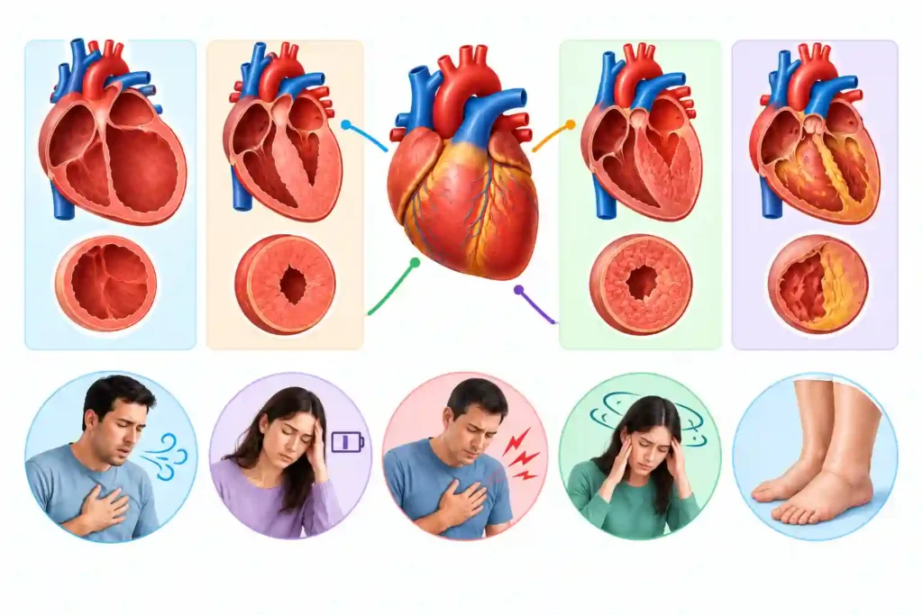
Generally, symptoms due to heart valve disease are shared with other heart diseases. Those symptoms are breathlessness, decreased stamina, fatigue, chest discomfort, dizziness, fainting, palpitations, and swelling in the legs or abdomen. Echocardiography is needed for diagnosing and assessing the affected valve and severity, because symptoms due to heart valve disease may be absent or nonspecific.
Mostly valve disease is found indirectly. Imagine a person who may blame slower walking on age; ankle swelling on a long day; or breathlessness on being out of shape. Meanwhile, in a routine checkup or for some other condition; a clinician may hear a murmur before the person feels anything unusual. But, sound alone cannot discover severity or anatomy.
Identifying heart valve disease symptoms can be helpful for you to notice a meaningful change and ask better questions, but it cannot determine a diagnosis. Some valve defects remain mild for years; others progress and put strain on the heart. Decisions are taken based on the valves involved, whether they are leaking or narrowed, their cause, symptoms, and objective test findings.
How Do Healthy Heart Valves Work?
There are four valves in the heart that keep blood moving forward through the heart: tricuspid, pulmonary, mitral, and aortic. Each opens to allow blood pass and closes to block backward flow. There may be a congenital valve defect (a valve can be affected from birth) or valve is affected later through aging, calcium buildup, infection, rheumatic disease, heart damage, or disorders of the valve tissue and supporting structures.
Valvular heart disease is an overarching term that is used for these problems. The burden of disease rises with age. A 2024 U.S. mortality analysis published in the Journal of the American Heart Association depicted that about 2.5% of adult prevalence has this disease. In 2025, the AHA informed that aortic stenosis is present in almost 12% of Americans aged 75 or older. Though estimates change with definitions and populations, both figures show the proportion of population involved and draw attention towards why new symptoms in older adults deserve assessment clinically.
What Is the Difference Between Stenosis and Regurgitation?
A diseased heart valve may be either stenosed or regurgitated. Difference is as follows:
- Stenosis implies that a valve opening has become narrowed or stiff, making it difficult for blood to move forward. The heart has to generate more pressure for pushing blood through.
- Regurgitation is sometimes also described as insufficiency or a leaky valve. It means that the valve does not close completely, permitting some blood to move backward.
Both problems can be mild, moderate, or severe. Both can enlarge or weaken heart chambers, thus contributing towards abnormal rhythms, or leading to heart failure over time. A valve may have both stenosis as well as regurgitation. Further, more than one valve can be affected. The label alone does not establish symptoms or treatment.
What Are Common Heart Valve Disease Symptoms?
As symptoms can develop slowly; therefore, changes are easiest to miss particularly when activity has silently decreased. Common patterns consist of:
- Shortness of breath during activity or while lying flat
- Decreased stamina, unusual fatigue, or weakness
- Chest pressure or discomfort, specifically with exertion
- Dizziness, light-headedness, fainting, or near-fainting
- Palpitations or a fast, pounding, or irregular heartbeat
- Swelling in the feet, ankles, legs, abdomen, or neck veins
- Speedy fluid-related weight gain or abdominal fullness
Aortic Stenosis Symptoms
The aortic valve controls blood leaving the left ventricle for the body. Age-related calcium buildup is a common cause of narrowing in older adults; a bicuspid valve present from birth can become diseased earlier. Rheumatic disease is another cause.
Typical aortic stenosis symptoms include exertional breathlessness, chest pressure, reduced exercise capacity, dizziness, or fainting. Heart-failure symptoms such as swelling or difficulty breathing when lying down may appear as disease advances. Symptoms in severe aortic stenosis are clinically important, but severity must be confirmed with testing rather than inferred from how dramatic a symptom feels.
Mitral Regurgitation Symptoms
The mitral valve lies between the left atrium and left ventricle. Regurgitation may result from changes in the valve leaflets, prolapse, infection, damage after a heart attack, or enlargement that pulls supporting structures apart.
Possible mitral regurgitation symptoms include breathlessness during activity or when lying down, fatigue, palpitations, cough, reduced exercise tolerance, and swelling. Chronic leakage may remain quiet while the heart compensates. Acute severe leakage can cause sudden breathlessness and low blood pressure and requires emergency care. An echocardiogram helps distinguish the mechanism and impact.
Does a Heart Murmur Mean Valve Disease?
A heart murmur is an extra sound produced by turbulent blood flow and heard with a stethoscope. Valve stenosis or regurgitation can cause a murmur, but not every murmur means structural valve disease. Innocent or flow murmurs can occur with a normal heart, including during pregnancy, fever, anemia, or increased blood flow.
Conversely, the loudness of a murmur does not consistently measure severity, and some significant valve problems produce subtle findings. A clinician considers timing, location, radiation, intensity, symptoms, pulse, and medical history. If structural disease is possible, echocardiography—not the sound alone—provides the essential information.
When Should Symptoms Be Treated as Urgent?
Call 911 in the United States, or your local emergency number, for severe or sudden breathlessness; chest pain or pressure; fainting or collapse; blue or gray lips; new confusion; signs of shock; or rapidly worsening symptoms. Do not drive yourself when emergency transport is available.
Fever with chills, new breathlessness, chest pain, or a new or changing murmur may raise concern for infective endocarditis, particularly in someone with a prosthetic valve, previous endocarditis, certain congenital conditions, or injection drug use. This needs prompt medical assessment rather than home treatment.
How Is Heart Valve Disease Checked?
Evaluation begins with symptoms, medical and family history, previous infections, pregnancy history, medicines, and known heart conditions. A clinician listens for murmurs, checks pulse and blood pressure, and looks for swelling or signs of heart failure. An electrocardiogram can identify rhythm changes or chamber strain, while a chest X-ray may show heart enlargement or lung congestion.
Echocardiography
An echocardiogram uses ultrasound to show valve anatomy, opening, closure, blood-flow direction and speed, chamber size, and pumping function. Doppler measurements help estimate narrowing or leakage. A standard transthoracic echocardiogram is performed from the chest. A transesophageal study places an ultrasound probe in the esophagus when closer images are needed; it requires specific preparation and carries different risks.
What Do Monitoring and Treatment Involve?
Mild valve disease may require periodic clinical visits and echocardiograms rather than immediate intervention. Monitoring intervals depend on the valve, severity, symptoms, and heart response. Report changes between visits instead of waiting for the next scheduled scan.
Medicines may manage blood pressure, fluid buildup, rhythm problems, or clot risk, but medication usually cannot mechanically open a severely narrowed valve or permanently correct major leakage. Some patients need valve repair or replacement through surgery or a catheter-based procedure. The timing can be important, including before symptoms become severe if tests show the heart is being affected.
The Practical Next Step
Pay attention to change, not simply symptom intensity. Record breathlessness, stamina, swelling, chest discomfort, fainting, and palpitations, then discuss the pattern with a clinician. Pursue emergency help for red flags. See where valve disease fits in Health Glow’s types-of-heart-disease pillar, and compare related heart-failure and cardiomyopathy guides.
Medical Disclaimer
This article provides general education and does not diagnose, treat, or replace individualized advice from a qualified healthcare professional. In an emergency, call your local