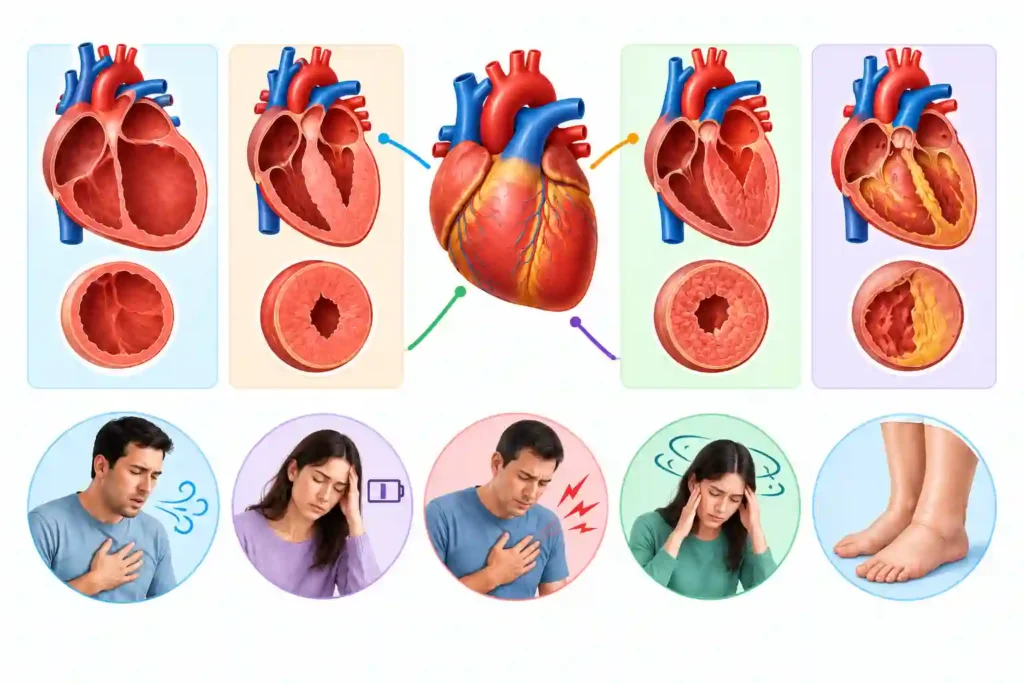
Cardiomyopathy confuses many readers and even technical people because the same word is used for hearts that are enlarged, abnormally thick, stiff, or part of their muscles is replaced by scar tissue. Cardiomyopathy exhibits differently in different people for example:
- One person may notice breathlessness;
- Another may have fainting, palpitations,
- Still some other people with cardiomyopathy may have no symptoms.
The appearance, cause, inheritance pattern, and rhythm risk can differ significantly; therefore, a label from a report needs clinical context.
Major categories of cardiomyopathy comprise dilated, hypertrophic, restrictive and arrhythmogenic cardiomyopathy. Symptoms can be breathlessness, fatigue, swelling, chest discomfort, palpitations, dizziness or fainting. However, some people do not have any symptoms and diagnosis needs cardiac evaluation.
If families understand cardiomyopathy types and symptoms; it will be helpful for them to organize questions without trying self-diagnosis. Cardiomyopathy is defined as a disease of heart muscle. It can damage pumping, filling, or electrical stability and may result in heart failure or arrhythmia.
What Are the Major Forms of Cardiomyopathy?
Mainly cardiomyopathy may manifest as:
- dilated heart chambers,
- hypertrophic heat muscles,
- restrictive, and arrhythmogenic.
Other categories of cardiomyopathy are left ventricular noncompaction and stress-induced cardiomyopathy. Classification of cardiomyopathy continues to change with upgrading in imaging and genetic knowledge, and also because some people have overlapping characteristics.
The American Heart Association estimates that almost 1 in 500 U.S. adults has hypertrophic cardiomyopathy (HCM). This figure could be more because many cases of cardiomyopathy remain undiagnosed. These statistics are applicable only to HCM, not cardiomyopathy as a whole. Prevalence assessments differ because mild disease can be quiet and diagnostic definitions keep on changing.
Dilated Cardiomyopathy
The term dilated cardiomyopathy applies to the condition where one or both ventricles become enlarged and the heart muscle contracts less successfully. The left ventricle is usually affected. Less effective pumping can decrease forward blood flow and let fluid to collect in the lungs, legs, or abdomen.
Causes of dilated cardiomyopathy may comprise inherited variants, viral or inflammatory injury, toxins like heavy alcohol exposure, certain chemotherapy drugs, nutritional or metabolic disorders, pregnancy-associated disease, and long-standing rapid rhythms. Sometimes no cause is detected. Symptoms may involve breathlessness, fatigue, swelling, reduced exercise capacity, palpitations, or dizziness.
Hypertrophic Cardiomyopathy
Hypertrophic cardiomyopathy (HCM) leads to abnormal thickening of heart muscle, repeatedly affecting the wall between the ventricles. Ventricle becomes stiff due to the thickening and thus weakening filling. HCM may be obstructive or nonobstructive. In obstructive HCM, muscle and mitral-valve motion narrow the path through which blood leaves the heart, while nonobstructive HCM has no major obstruction while resting.
HCM is generally inherited, but disease appearance differs even within a family. Possible symptoms incorporate breathlessness during exertion, chest discomfort, palpitations, dizziness, or fainting. Some people remain asymptomatic, i.e. disease remains silent. HCM can raise the risk of atrial fibrillation and dangerous ventricular rhythms in a smaller group. Specific risk cannot be evaluated from wall thickness or symptoms alone.
Restrictive Cardiomyopathy
Restrictive cardiomyopathy denotes the condition where the ventricles become stiff and are unable to relax or fill normally, even when pumping strength seems preserved at early stage. Resultantly, pressure backs up into the atria, lungs, and veins, creating breathlessness, swelling, fatigue, or abdominal fullness.
Possible causes of restrictive cardiomyopathy include amyloidosis, sarcoidosis, iron overload, radiation injury, certain connective-tissue or metabolic disorders, and inherited conditions. The presentation of disease can resemble constrictive pericarditis, valve disease, or heart failure with preserved ejection fraction; therefore, cautious imaging and testing are important.
Arrhythmogenic Cardiomyopathy and Other Forms
Arrhythmogenic cardiomyopathy manifests when heart muscle are replaced by fibrous or fatty tissue. It frequently affects the right ventricle but sometimes the left or both. Mostly, it is inherited and can lead to palpitations, fainting, ventricular arrhythmias, or abrupt cardiac arrest.
Congenital cardiomyopathy called left ventricular noncompaction has prominent muscular ridges and deep spaces in the ventricular wall; interpretation needs expertise because similar imaging features can appear without disease. Cardiomyopathy induced by stress results in sudden, usually temporary, weakness after intense physical or emotional stress and can give impression of a heart attack. Pregnancy-associated cardiomyopathy progresses late in pregnancy or in the months after delivery.
What Symptoms Can Cardiomyopathy Cause?
Usual cardiomyopathy symptoms consist of:
- Shortness of breath during activity, at rest, or while lying flat
- Unusual fatigue, weakness, or declining exercise tolerance
- Swelling in the feet, ankles, legs, abdomen, or neck veins
- Chest pressure or discomfort
- Palpitations, racing, skipped beats, or an irregular pulse
- Dizziness, light-headedness, fainting, or near-fainting
- Cough, rapid fluid-related weight gain, or reduced appetite
When Do Symptoms Need Urgent Help?
Call 911 in the United States, or your local emergency number,
Symptoms of cardiomyopathy that need urgent help include:
- chest pain or pressure;
- severe or sudden breathlessness;
- fainting or collapse;
- a sustained rapid or irregular heartbeat with weakness or dizziness;
- blue or gray lips;
- new confusion;
- signs of stroke.
When you come across above conditions, call 911 in the United States, 1122 in Pakistan or your local emergency number. Meanwhile, provide first aid to the patient. If someone is unresponsive and not breathing normally, start CPR and you may use an automated external defibrillator provided it is available and you are able to use it.
Fainting during exercise, specifically with a family history of cardiomyopathy or sudden unexplained death, should be assessed urgently. Prompt care is also necessary in case of sudden symptoms during pregnancy or the months after delivery. Those symptoms include breathlessness at rest, chest pain, fainting, or rapidly increasing swelling. Avoid driving yourself when emergency transport is available.
Can Cardiomyopathy Be Inherited?
Yes, several cardiomyopathies can be due to inherited genetic variants, specifically hypertrophic and arrhythmogenic forms and some dilated or restrictive cases. An inherited variant does not expect same symptoms or severity in every relative. Some carriers may develop disease at a later age; it could be mild, or not at all.
Once an inherited form is suspected or verified, genetic counseling can explain the advantages, limitations, and potential results of testing. First-degree relatives should be offered clinical screening, like an ECG and echocardiogram, with timing determined by age, family findings, and professional guidance. A direct-to-consumer result should never be be used in silo to diagnose disease or clear someone from follow-up.
How is Cardiomyopathy Diagnosed?
Evaluation A combination of symptoms, examination, personal history, family history, and tests are utilized for assessment and diagnosing cardiomyopathy. Clinicians also assess blood pressure, pregnancy history, infections, alcohol or drug exposure, medicines, chemotherapy or radiation, and systemic conditions. Blood tests may assess kidney, liver, thyroid, iron, inflammation, or markers of cardiac strain or injury.
An ECG judges rhythm and electrical patterns. Echocardiography confirms wall thickness, chamber size, pumping, filling, valves, and possible obstruction. Cardiac MRI presents detailed muscle structure and can identify inflammation or scar. Rhythm monitors capture intermittent arrhythmias, while exercise testing may assess symptoms, blood-pressure response, obstruction, or rhythm under controlled conditions.
How Is Cardiomyopathy Managed?
Management of cardiomyopathy is not simple, and it is based on subtype, cause, symptoms, pumping and filling function, obstruction, and rhythm or clot risk. Management may incorporate medicines, treatment of an underlying condition, activity planning, rhythm monitoring, catheter ablation, a pacemaker or defibrillator, surgery for selected obstructive HCM, or advanced heart-failure therapies.
Advice about competitive sports or rigorous exercise should be tailored for individuals. Diagnosis alone does not determine what activity is safe, and blanket restriction can also be harmful. A cardiology team should think about subtypes, symptoms, rhythm history, imaging, family history, treatment, and personal goals.
People with cardiomyopathy benefit from a clear follow-up schedule and action plan. Take medicines as instructed, discuss pregnancy planning early, do not take unreviewed supplements or stimulants, and report important changes in breathing, swelling, fainting, chest discomfort, or palpitations.
The Practical Next Step
If cardiomyopathy is suspected, record symptoms and assemble a detailed family heart history before the appointment. Seek emergency help for red flags. Compare cardiomyopathy with heart failure, arrhythmia, and other conditions in Health Glow’s types-of-heart-disease pillar, then use the related cluster guides to prepare questions.
Medical Disclaimer
This article provides general education and does not diagnose, treat, determine exercise eligibility, or replace individualized advice from a qualified healthcare professional. In an emergency, call your local emergency number.